Skin Worksheet Answers WikiEducator
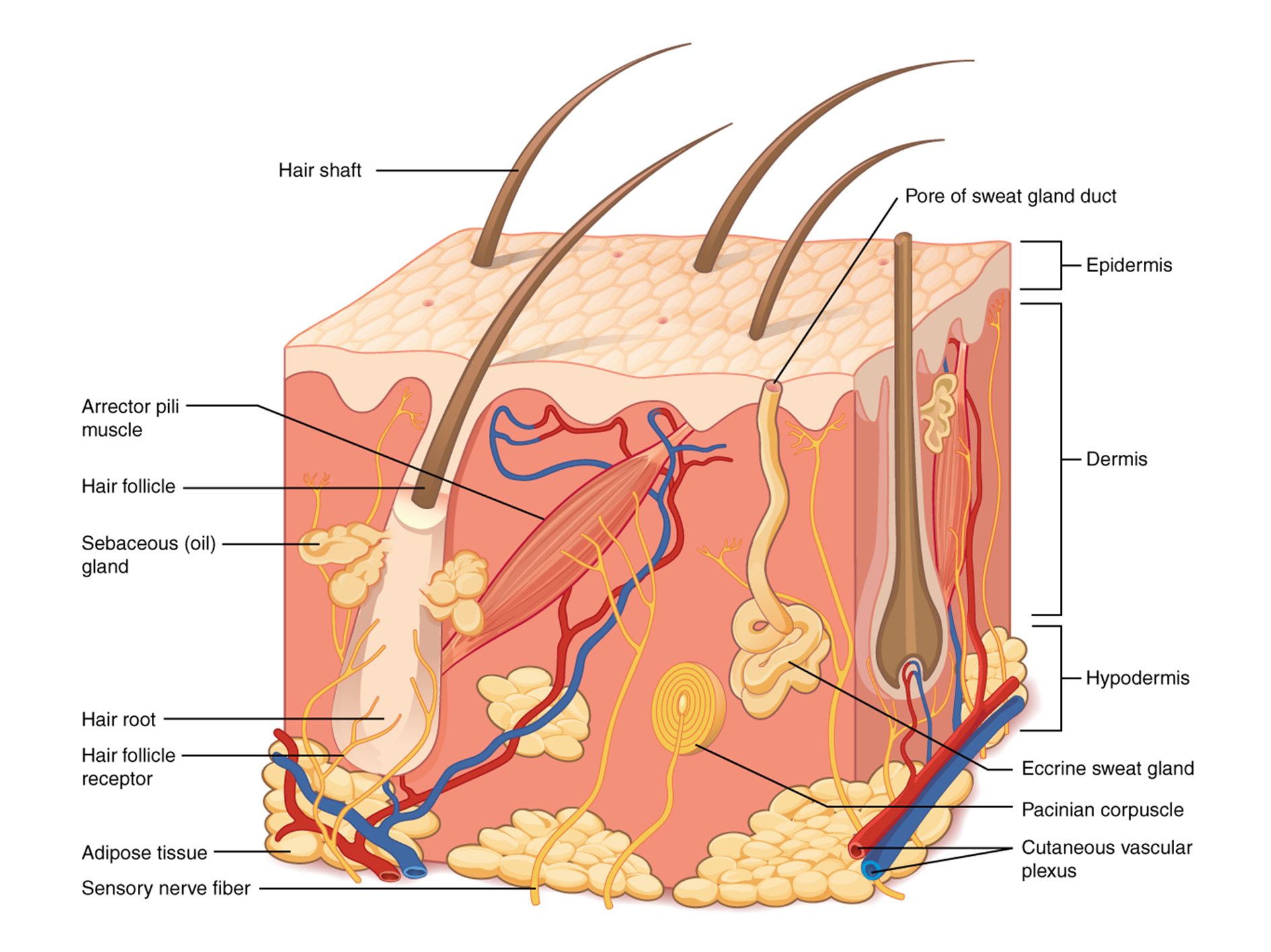
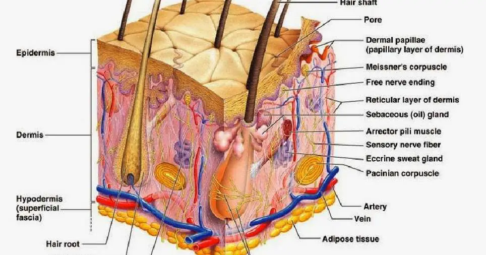
Figure 5.2 Layers of Skin The skin is composed of two main layers: the epidermis, made of closely packed epithelial cells, and the dermis, made of dense, irregular connective tissue that houses blood vessels, hair follicles, sweat glands, and other structures. Beneath the dermis lies the hypodermis, which is composed mainly of loose connective.

Structure and composition of the skin [5]. Download Scientific Diagram
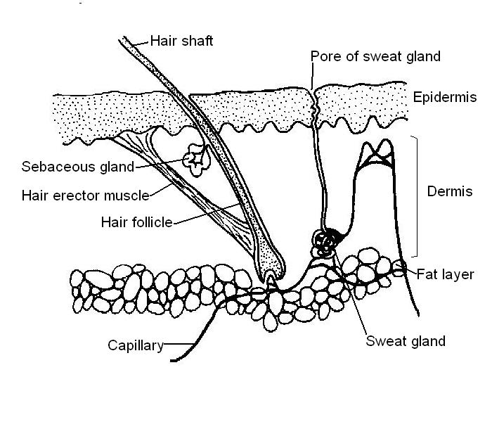
Printout. The skin is an organ that forms a protective barrier against germs (and other organisms) and keeps the inside of your body inside your body, and keeps what's outside of your body outside. Skin also helps maintain a constant body temperature. Human skin is only about 0.07 inches (2 mm) thick. Skin is made up of two layers that cover a.

Diagram of human skin layers Charlotte Desire
Skin is the largest organ of the body. It has an area of 2 square metres (22 square feet) in adults, and weighs about 5 kilograms. The thickness of skin varies from 0.5mm thick on the eyelids to 4.0mm thick on the heels of your feet. Skin is the major barrier between the inside and outside of your body!
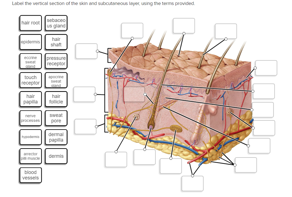
Solved Label The Vertical Section Of The Skin And Subcuta...
This Osmosis High-Yield Note provides an overview of Skin Structures essentials. All Osmosis Notes are clearly laid-out and contain striking images, tables, and diagrams to help visual learners understand complex topics quickly and efficiently. Find more information about Skin Structures: Skin anatomy and physiology. Hair, skin and nails.

Blister Care
Skin Diagram. The largest organ in the human body is the skin, covering a total area of about 1.8 square meters. The skin is tasked with protecting our body from external elements as well as microbes. The skin is also responsible for maintaining our body temperature - this was apparent in victims who were subjected to the medieval torture of.

human skin cells labeled Google Search Subcutaneous tissue, Skin
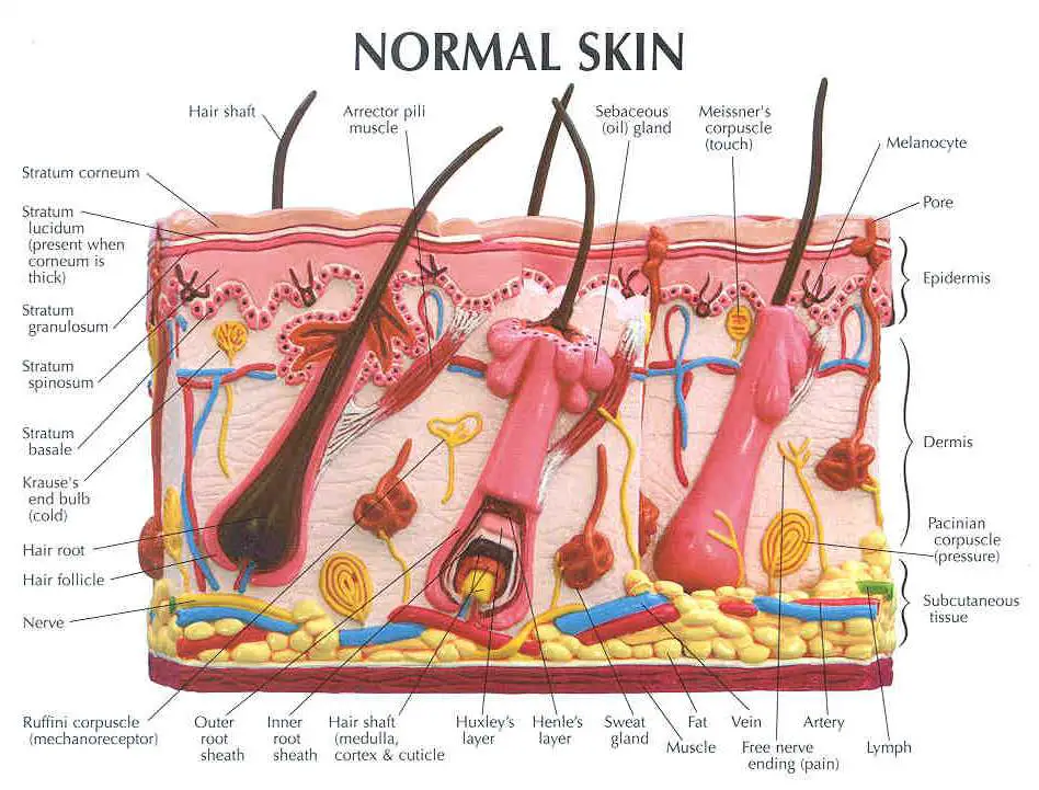
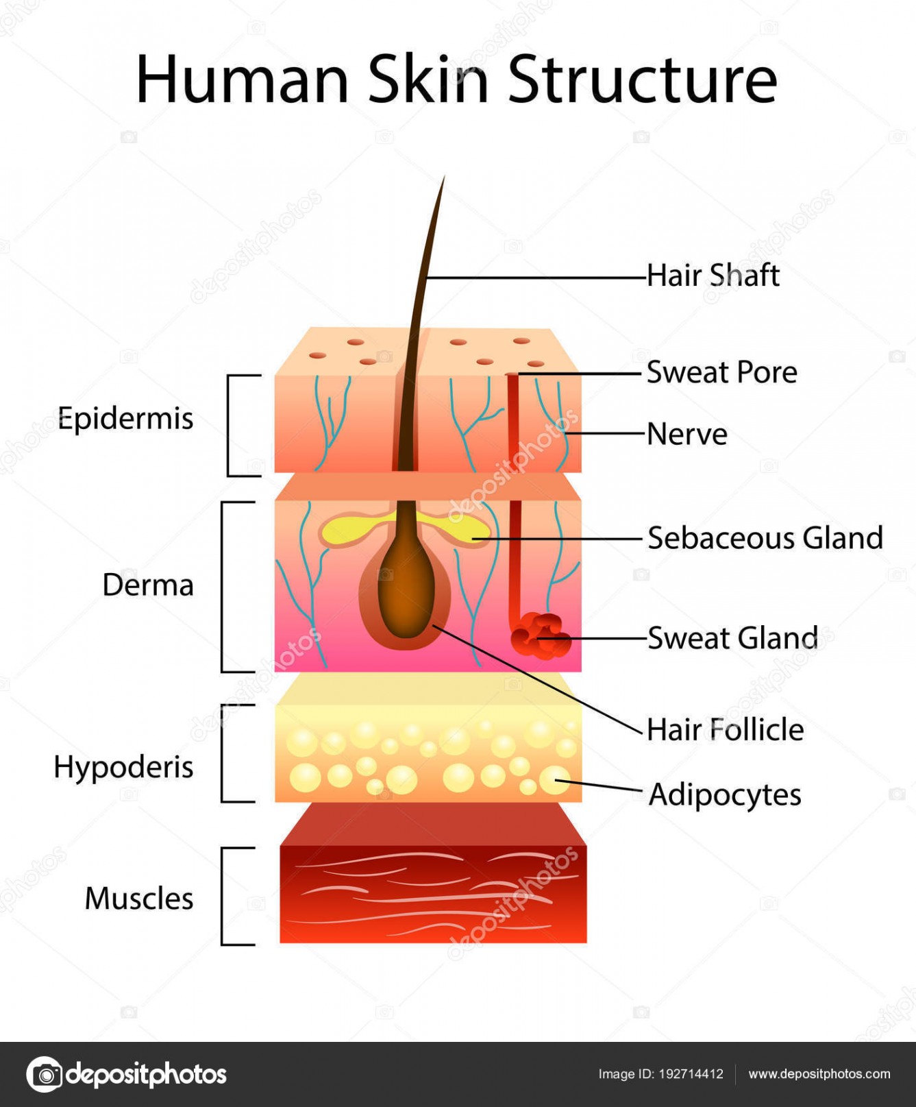
The skin has three basic layers, each with a different role. The number of skin layers that exists depends on how you count them. You have three main layers of skin—the epidermis , dermis, and hypodermis (subcutaneous tissue). Within these layers are additional layers. If you count the layers within the layers, the skin has eight or even 10.
Skin diagram labeled
Functions. The skin has a significant capacity for renewal and crucial roles for the normal functioning of the human body. It is an effective barrier against potential pathogens and protects against mechanical, chemical, osmotic, thermal and ultraviolet radiation damage (through melanin). The skin also takes part in a variety of biochemical synthetic processes, such as vitamin D production.

35 Label Skin Diagram Labels Design Ideas 2020
Skin is the largest organ in the body and covers the body's entire external surface. It is made up of three layers, the epidermis, dermis, and the hypodermis, all three of which vary significantly in their anatomy and function. The skin's structure is made up of an intricate network which serves as the body's initial barrier against pathogens, UV light, and chemicals, and mechanical injury.

Human Anatomy Diagrams To Label koibana.info Skin anatomy, Human
The skin is the body's largest organ. It serves many important functions, including. Protecting the body against trauma. Regulating body temperature. Maintaining water and electrolyte balance. Sensing painful and pleasant stimuli. Participating in vitamin D synthesis. The skin keeps vital chemicals and nutrients in the body while providing a.
POSTECH University develops 3D bioprinting technique that grows human
'Skin Diagram || How to draw and label the parts of skin' is demonstrated in this video tutorial step by step.The sense of touch had received supreme importa.
Skin diagram labeled
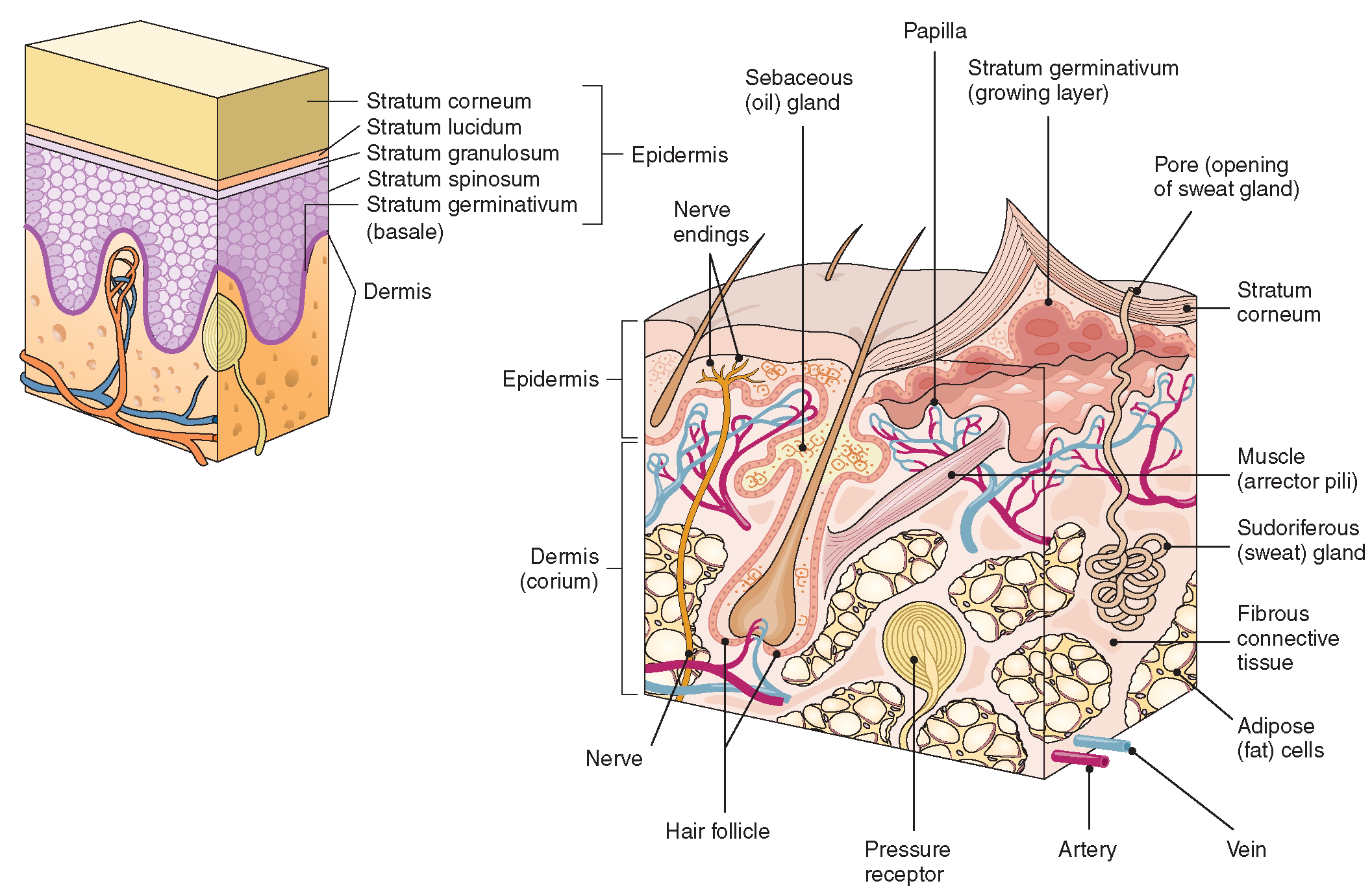
Skin that has four layers of cells is referred to as "thin skin.". From deep to superficial, these layers are the stratum basale, stratum spinosum, stratum granulosum, and stratum corneum. Most of the skin can be classified as thin skin. "Thick skin" is found only on the palms of the hands and the soles of the feet.
The Integumentary System (Structure and Function) (Nursing) Part 1
Diagram of human skin structure. Image. Add to collection. Rights: The University of Waikato Te Whare Wānanga o Waikato Published 1 February 2011 Size: 100 KB Referencing Hub media. The epidermis is a tough coating formed from overlapping layers of dead skin cells.

Science Homework, Skin Drawing, Human Body Anatomy, Sense Of Touch
The Layers of Your Skin. Your skin includes three layers known as epidermis, dermis, and fat. Some health issues, such as dermatitis and infections, can affect how these different layers work to.

Quia Class Page 5th Period
The skin is composed of two main layers: the epidermis, made of closely packed epithelial cells, and the dermis, made of dense, irregular connective tissue that houses blood vessels, hair follicles, sweat glands, and other structures. Beneath the dermis lies the hypodermis, which is composed mainly of loose connective and fatty tissues.
Labelled Pictures Of Human Skin / skin diagram /medical/anatomy/skin
The skin is the body's largest and primary protective organ, covering its entire external surface and serving as a first-order physical barrier against the environment. Its functions include temperature regulation and protection against ultraviolet (UV) light, trauma, pathogens, microorganisms, and toxins. The skin also plays a role in immunologic surveillance, sensory perception, control of.
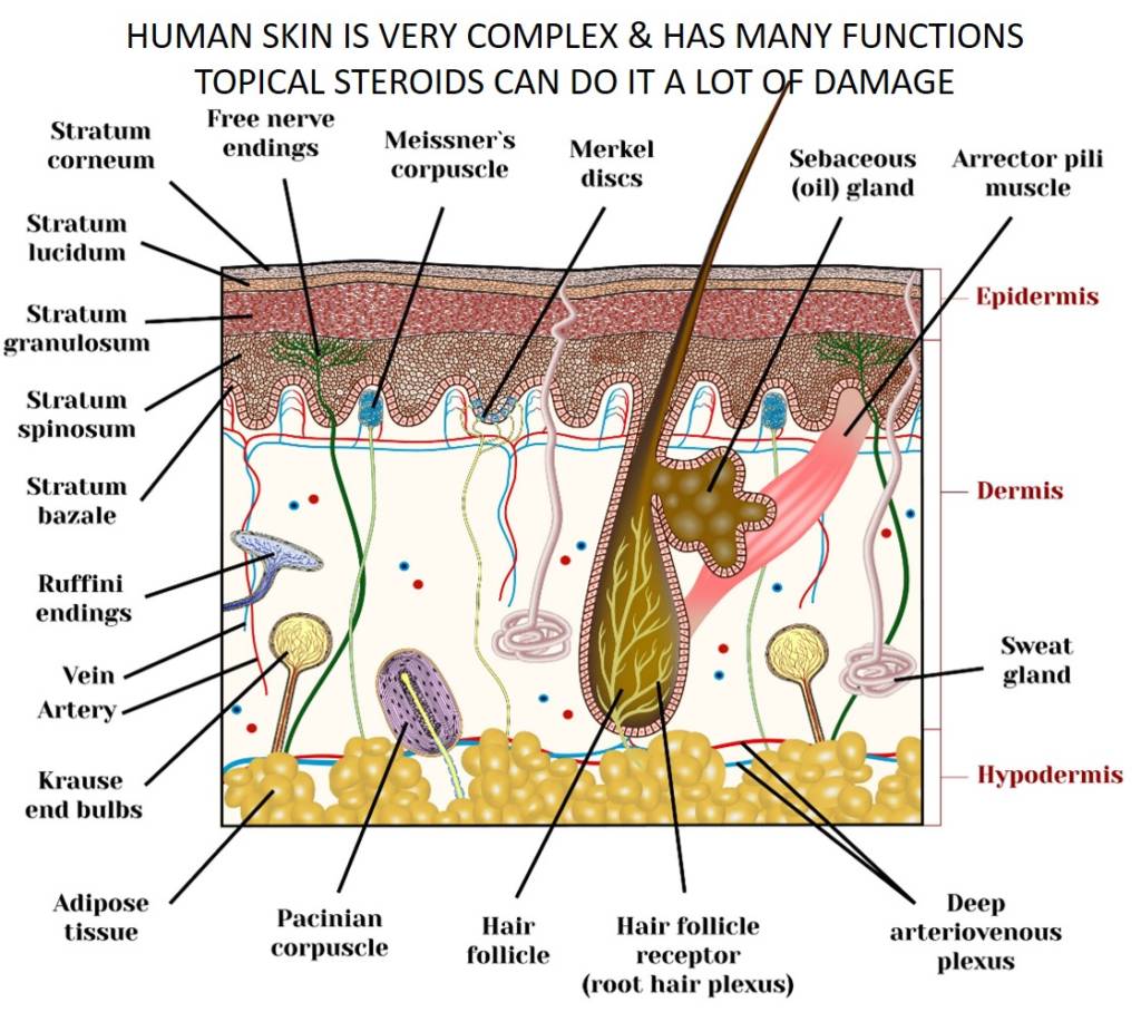
Skin topical steroid withdrawal with wheatgrass extract A
Facts about the skin. The skin is the body's largest organ. It covers the entire body. It serves as a protective shield against heat, light, injury, and infection. The skin also: Regulates body temperature. Stores water and fat. Is a sensory organ. Prevents water loss. Prevents entry of bacteria. Acts as a barrier between the organism and its.